A glycosidic bond is a covalent bond formed between the anomeric (hemiacetal) carbon of a carbohydrate and a nucleophilic atom, typically an oxygen (O), nitrogen (N) or sulphur (S) atom, from another molecule that carries out the attack.[1]
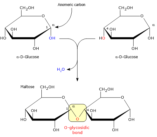
The reaction leading to the formation of the glycosidic bond is a condensation reaction, which involves the release of a water molecule and the formation of an acetal.[2]
The glycosidic bond can be classified according to at least three main criteria: the atom carrying out the nucleophilic attack, the initial configuration of the carbon in the hemiacetal group (α or β), and the position of the carbon atoms involved in the bond. This latter classification is particularly useful for understanding the structure of disaccharides, oligosaccharides and polysaccharides, as well as for clarifying which carbohydrates can be hydrolysed and digested in the human intestine.[3][4]
Summary: Key Points
- Definition and chemistry: a covalent bond between the hemiacetal carbon of a molecule and a nucleophile, converting the hemiacetal into an acetal with the release of a water molecule.
- Classification by atom type: classified according to the nucleophilic atom into O-glycosidic, N-glycosidic, and S-glycosidic bonds.
- Anomeric classification: originates from the nucleophilic attack on either face of the sp2 hybridized (planar) carbonyl carbon, yielding α and β anomers.
- Classification by linkage position: includes linear (1→4) bonds, (1→6) branch points, and linkages between anomeric carbons such as α-(1→2)β in sucrose and α,α-(1→1) in trehalose.
- Nutritional impact and digestion: humans possess enzymes capable of hydrolyzing α-linkages, with the notable exception of the β-(1→4) linkage in lactose, which is cleaved by lactase.
Contents
- O-, N– and S-glycosidic bonds
- α and β glycosidic bonds
- Glycosidic bond and position of the involved atoms
- References
O-, N– and S-glycosidic bonds
Depending on the nature of the nucleophilic atom (oxygen, nitrogen or sulphur) that attacks the anomeric carbon in the hemiacetal, glycosidic bonds are classified into three main categories: O-, N– and S-glycosidic bonds, the latter being the rarest.[1]
O-glycosidic bond
The O-glycosidic bond is the most common form found in nature. It is formed when the nucleophile is the oxygen of a hydroxyl group (−OH). This covalent bond is fundamental to the formation of disaccharides, the most common of which in the human diet are sucrose and lactose, oligosaccharides and storage polysaccharides, such as starch in plants and glycogen in animals, or structural polysaccharides, such as cellulose.
It is also found in O-linked glycoproteins, where the sugar binds to the oxygen of the hydroxyl group on the side chain of amino acids such as serine or threonine.[4]
N-glycosidic bond
An N-glycosidic bond is formed when the nucleophile carrying out the attack is the nitrogen of an amino group (−NH2 or −NH−). This bond plays a crucial biological role: it links nitrogenous bases, that is, purines and pyrimidines, to ribose or deoxyribose in the structure of nucleotides and nucleic acids, namely DNA and RNA. It is also found in N-linked glycoproteins, in which the sugar ring is bound to the amide nitrogen of the asparagine side chain.[5]
S-glycosidic bond
Of the three types of glycosidic bond, the S-glycosidic bond is the rarest and is formed when the nucleophilic attack is carried out by the sulphur atom of a thiol group (−SH). It is characteristic of glucosinolates, secondary metabolites found mainly in the Brassicaceae, such as broccoli and cabbage, where they are involved in plant defence mechanisms against herbivores.[6]
| Atomic category | Nucleophilic atom and bond type | Geometry and position | Example substrates/molecules | Chemical and structural features |
|---|---|---|---|---|
| O-glycosidic | O-glycosidic (monocyclic acetal bond) | Linear α-(1→4) and β-(1→4) | Amylose, cellulose, maltose, lactose | Oxygen bridge between C1 and C4. Anomeric orientation (α helical vs β linear/extended) dictates three-dimensional structure and solubility. |
| O-glycosidic (branching bond) | Branched α-(1→6) | Amylopectin, glycogen, isomaltose | Oxygen bridge between anomeric C1 and primary C6 outside the ring, imparting high structural flexibility. | |
| O-glycosidic (inter-anomeric bond) | α-(1→2)β and α,α-(1→1) | Sucrose, trehalose | Direct linkage between the anomeric carbons of two monosaccharides. Eliminates free hemiacetal groups, yielding a non-reducing sugar. | |
| N-glycosidic | N-glycosidic (amine/nucleosidic bond) | β-N-glycosidic between anomeric C1 and nitrogen (N9/N1) | Nucleosides, nucleotides (DNA, RNA), NAD+, glycoproteins | Nitrogen bridge between C1 of ribose/deoxyribose and heterocyclic nitrogen of purine (N9) or pyrimidine (N1) bases or peptide side chains (asparagine). |
| S-glycosidic | S-glycosidic (thioacetal bond) | β-S-glycosidic between anomeric C1 and thiol group | Glucosinolates (sinigrin, glucoraphanin) | Sulphur bridge linked to the anomeric carbon of D-glucose. Characteristic of secondary metabolites in Brassicaceae, hydrolyzed by the myrosinase enzyme. |
α and β glycosidic bonds
Depending on the initial configuration of the carbon atom in the hemiacetal group, the glycosidic bond may be an α or a β bond.
During the intramolecular cyclisation that closes the monosaccharide ring, the carbon of the carbonyl group, which has sp2 hybridisation and is planar, undergoes nucleophilic attack by the internal hydroxyl group.
Since the attack can occur on either side of the carbonyl plane, this carbon becomes a new centre of chirality, known as the anomeric carbon, giving rise to two distinct stereoisomers, termed the α and β anomers.
- α-Isomer: the anomeric hydroxyl group is in the trans position relative to the reference group, i.e. the −CH2OH group in D-sugars, oriented downwards in the Haworth projection.
- β-Isomer: the anomeric hydroxyl group is in the cis position relative to the reference group, oriented upwards in the Haworth projection. The β configuration is often the thermodynamically favoured form.
In aqueous solution, free monosaccharides undergo mutarotation, that is, a dynamic equilibrium between the open form and the two α and β anomers.[1][2]
Glycosidic bond and position of the involved atoms
The position of the participating atoms, the nature of the bridging atom (O, N, S), and the anomeric configuration (α or β) dictate both the spatial geometry of the molecules and their susceptibility to hydrolysis by human digestive enzymes or the gut microbiota. Based on these features, standard α-(1→4), β-(1→4), and α-(1→6) linkages, inter-anomeric α-(1→2)β and α,α-(1→1) bonds, linkages characteristic of prebiotic and structural fibers such as β-(1→3), as well as the heteroatom linkages (N– and S-glycosidic) are identified.[2]
| Category | Glycosidic bond type | Substrate | Main hydrolytic enzyme | Human digestive site and bioaccessibility |
|---|---|---|---|---|
| α-Linkages | α-(1→4) and α-(1→6) | Starch, glycogen, maltose | α-Amylase (EC 3.2.1.1), maltase-glucoamylase (EC 3.2.1.20), isomaltase (EC 3.2.1.10) | Mouth and small intestine (fully digestible/rapidly absorbed as glucose) |
| α-(1→2) | Sucrose | Sucrase-isomaltase (EC 3.2.1.48 and 3.2.1.10) | Brush border of the small intestine (fully digestible/hydrolyzed into glucose + fructose) | |
| α,α-(1→1) | Trehalose | Trehalase (EC 3.2.1.28) | Brush border of the small intestine (digestible/hydrolyzed into 2 glucose molecules; fermented in case of enzyme deficiency) | |
| β-Linkages | β-(1→4) [galactose-glucose] | Lactose | Lactase (EC 3.2.1.108) | Brush border of the small intestine (digestible in individuals with lactase persistence; fermented in case of deficiency) |
| β-(1→4) [glucose-glucose] | Cellulose | Cellulase (EC 3.2.1.4; absent in humans) | Colon/Gut microbiota (indigestible dietary fiber; partially fermented by anaerobic bacteria into short-chain fatty acids) | |
| β-(1→3) and β-(1→4)/β-(1→6) | β-Glucans, inulin, hemicellulose | Carbohydrate-active bacterial enzymes (Glycoside hydrolases; EC 3.2.1) | Colon/Gut microbiota (prebiotic dietary fiber; fermented into short-chain fatty acids) | |
| Heteroatom linkages | S-Glycosidic and N-glycosidic | Glucosinolates, nucleosides | Myrosinase (EC 3.2.1.147; plant/microbial), nucleosidases | Upper gastrointestinal tract and gut microbiota (metabolized into bioactive isothiocyanates or free purines/pyrimidines) |
α-(1→4) vs β-(1→4) glycosidic bond
The α-(1→4) bond is a linear bond that links D-glucose units in glycogen as well as in amylose, which, together with amylopectin, is one of the two components of starch. Mammals synthesise the enzyme α-amylase, which is capable of specifically hydrolysing this bond to release glucose for energy.[4]
The β-(1→4) bond is characteristic of cellulose, the main structural polysaccharide in plants. Due to the β orientation, the chains adopt a linear and rigid conformation, stabilised by interchain hydrogen bonds. Mammals lack the enzyme β-glucosidase (EC 3.2.1.21) and cannot hydrolyse this bond. This makes cellulose a fibre; it is therefore indigestible but can nevertheless be fermented by the gut microbiota.[7]
However, most mammals are able to hydrolyse the β-(1→4) glycosidic bond that links galactose and glucose in the disaccharide lactose. This is made possible by the presence, on the brush border of enterocytes, of the enzyme lactase, a β-galactosidase specific to the β-(1→4) glycosidic bond in lactose.[8]
α-(1→6) glycosidic bond
The α-(1→6) bond forms the branching points in the structures of amylopectin and glycogen. Since α-amylase primarily hydrolyses (1→4) bonds, the cleavage of the (1→6) branches requires a specific enzyme, the debranching enzyme isomaltase.[4]
(1→2) and (1→1) glycosidic bonds between anomeric carbons
When the glycosidic bond directly involves the anomeric carbons of both monosaccharides, as in (1→2) and (1→1) bonds, the molecule loses its free hemiacetal groups and becomes a non-reducing sugar.[1]
An example of a bond between anomeric carbon atoms is the α-(1→2)β bond in sucrose, where the anomeric carbon C1 of α-D-glucose bonds to the anomeric carbon C2 of β-D-fructose. By involving both the hemiacetal groups responsible for the reducing power, sucrose becomes a non-reducing sugar. In humans, this bond is effectively hydrolysed by sucrase-isomaltase, making the two monosaccharides immediately available for absorption.[4]
The α,α-(1→1) bond, found in trehalose, is formed between the two anomeric C1 carbons of two α-D-glucose molecules; this too blocks reactivity and renders the sugar non-reducing. In humans, this bond is cleaved by the enzyme trehalase.[5]
In summary, the stereochemical specificity of digestive enzymes, capable of recognising the α geometry but not the β geometry in (1→4) carbohydrate chains, with the sole exception of lactose, establishes the boundary between what constitutes an energy source for humans and what constitutes dietary fibre.[9]
References
- a b c d Soderberg T. Organic chemistry with a biological emphasis. Volume I. Chemistry Publications. 2019.
- a b c Solomons T.W.G., Fryhle C.B., Snyder S.A. Solomons’ organic chemistry. 12th Edition. John Wiley & Sons Incorporated, 2017.
- ^ IUPAC-IUBMB Joint Commission on Biochemical Nomenclature (JCBN). Nomenclature of carbohydrates. Recommendations 1996. Eur J Biochem 1997;243:9-9. doi:10.1111/j.1432-1033.1997.09_1a.x
- a b c d e Nelson D.L., Cox M.M. Lehninger. Principles of biochemistry. 8th Edition. W.H. Freeman and Company, 2021.
- a b Berg J.M., Tymoczko J.L., Gatto G.J., Stryer L. Biochemistry. 9th Edition. W.H. Freeman and Company, 2019.
- ^ Halkier B.A., Gershenzon J. Biology and biochemistry of glucosinolates. Annu Rev Plant Biol 2006;57:303-33. doi:10.1146/annurev.arplant.57.032905.105228
- ^ Lattimer J.M., Haub M.D. Effects of dietary fiber and its components on metabolic health. Nutrients 2010;2(12):1266-89. doi:10.3390/nu2121266
- ^ Zecca L., Mesonero J.E., Stutz A., Poirée J.C., Giudicelli J., Cursio R., Gloor S.M., Semenza G. Intestinal lactase-phlorizin hydrolase (LPH): the two catalytic sites; the role of the pancreas in pro-LPH maturation. FEBS Lett 1998;435(2-3):225-8. doi:10.1016/s0014-5793(98)01076-x
- ^ Guyton A.C., Hall J.E. Textbook of medical physiology. 14th Edition. Philadelphia: Elsevier, 2021.
Domande Frequenti
From a chemical point of view, what is a glycosidic bond and how is it formed?
It is a covalent bond joining a carbohydrate's anomeric carbon to another molecule. Formed via condensation, a nucleophilic attack by a hydroxyl, amine, or thiol group leads to the elimination of a water molecule.
What is the structural difference between α and β bonds?
It lies in the oxygen orientation at the anomeric C1 carbon. The α isomer has the oxygen in trans relative to the −CH2OH group (digestible by amylase), while the β isomer has it in cis, imparting structural rigidity and resistance to human digestive enzymes (except in lactose).
Why can humans digest plant starch but cannot digest cellulose?
Humans secrete α-amylase to cleave α-(1→4) bonds in starch, but lack cellulase for β-(1→4) bonds in cellulose. Thus, cellulose acts as indigestible dietary fiber, partially fermented by the gut microbiota in the colon.
What are the key differences between O-, N-, and S-linkages?
Classification depends on the nucleophilic atom: oxygen forms O-glycosidic bonds (starch, cellulose), nitrogen creates N-glycosidic bonds (nucleotides in DNA/RNA), and sulfur yields S-glycosidic bonds (glucosinolates in Brassicaceae)