Disaccharides are carbohydrates consisting of two monosaccharides joined by a covalent bond known as a glycosidic bond.[1]
Disaccharides found in the human diet include sucrose, lactose, maltose and trehalose, which are structural isomers that differ in solubility, sweetening power and chemical reactivity.[2]
They are digested in the mucosa of the small intestine, a process that requires the action of a specific set of glycosidases located on the brush border of the enterocytes.[3] These enzymes exhibit high stereospecificity, distinguishing between the α and β configurations of the glycosidic bond.[4]
When the activity of one or more of these enzymes is absent or deficient, due to primary genetic abnormalities or secondary damage to the intestinal mucosa, malabsorption of disaccharides occurs. The undigested disaccharide proceeds to the colon, where it acts as an osmotically active solute and is fermented by the gut microbiota, triggering gastrointestinal symptoms.[5][6]
Summary: Key Points
- Chemical properties: disaccharides are isomers formed by two monosaccharides linked by an O-glycosidic bond. They are classified as reducing (lactose and maltose) and non-reducing (sucrose and trehalose) based on the presence of a free hemiacetal group.
- Stereospecificity of anomers: the closure of the monosaccharide ring creates α and β anomers. The geometry of the resulting glycosidic bond requires stereospecific hydrolytic enzymes.
- Intestinal digestion: the glycosidases of the brush border (sucrase-isomaltase, maltase-glucoamylase, trehalase and lactase) selectively hydrolyse the glycosidic bonds of dietary disaccharides. Lactase is the only digestive β-glycosidase in mammals.
- Pathophysiology of enzyme deficiency: a deficiency in glycosidases leads to the intraluminal accumulation of the disaccharide, causing it to be fermented by the gut microbiota and resulting in osmotic-fermentative diarrhoea and gastrointestinal disturbances.
- Aetiology of deficiencies: deficiencies may be congenital or secondary and transient, resulting from diseases or damage to the intestinal epithelium.
Contents
- Chemical properties
- Glycosidic bonds in disaccharides
- Dietary disaccharides
- Digestion of disaccharides and glycosidases
- References
Chemical properties
Disaccharides are organic molecules formed by the condensation of two monosaccharides, joined by a covalent bond known as a glycosidic bond.[1]
Sucrose, lactose, maltose, and trehalose have the general chemical formula C12H22O11 and a molecular weight of approximately 342.30 g/mol, differing only in the arrangement of their atoms; they are therefore isomers.[7]
Depending on whether or not they contain a free hemiacetal group capable of acting as a reducing aldehyde group, disaccharides can be reducing or non-reducing. Lactose and maltose belong to the former group, whilst sucrose and trehalose belong to the latter, as the hemiacetal group of the second monosaccharide is also involved in the formation of the glycosidic bond.[2]
Lactose and maltose, being reducing disaccharides and therefore possessing a free hemiacetal group, can undergo the Maillard reaction; during cooking, they react with the amino acids in proteins, leading to the formation of aromatic compounds and the browning of food surfaces during thermal processing.[8]
Disaccharides, like monosaccharides, are molecules that are readily soluble in water and taste sweet. Their sweetness is measured relative to that of sucrose, which is conventionally assigned a value of 1. Lactose has a value of 0.16–0.4, maltose of 0.3–0.5, and trehalose of approximately 0.45.[9]
Glycosidic bonds in disaccharides
In disaccharides, the two monosaccharides are joined by a glycosidic bond, a covalent bond formed between the anomeric carbon of a hemiacetal group in one monosaccharide and a nucleophilic atom, typically an oxygen, nitrogen, or sulphur atom, belonging to another molecule. The reaction leading to the formation of the glycosidic bond is a condensation reaction that involves the loss of a water molecule and results in the formation of an acetal.
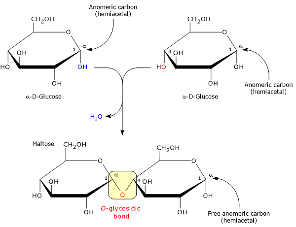
In disaccharide synthesis, the attacking nucleophilic atom belongs to a second monosaccharide, forming an O-glycosidic bond.[8]
Stereospecificity of the glycosidic bond
Depending on the initial configuration of the hemiacetal carbon, the glycosidic bond can be of the α or β type.[4]
If we consider the hemiacetal carbon of any monosaccharide, it exhibits the phenomenon of anomerism: in the closed-ring cyclic form of the molecule, two distinct stereochemical configurations are possible, referred to as α or β anomers.
During the intramolecular cyclisation that leads to ring closure, with the conversion of the sp2 carbonyl carbon into a chiral centre:
- the α isomer is formed when the anomeric hydroxyl group is in the trans position relative to the stereocentric reference group, i.e. oriented downwards in the Haworth projection for the D series;
- the β isomer is formed when the anomeric hydroxyl group is in the cis position relative to the reference group, i.e. oriented upwards in the Haworth projection for the D series.
The β configuration is generally the thermodynamically predominant form in aqueous solution.[1][8]
Dietary disaccharides
The most common disaccharides in the human diet are sucrose and lactose. Trehalose is less common, whilst maltose, unless used as an additive, is derived almost exclusively from the hydrolysis of starch, specifically the amylose and amylopectin that make it up, by the action of α-amylase.[7]
| Disaccharide | Constituent monosaccharides | Type of glycosidic bond | Hemiacetal group and reducing power | Relative sweetness (sucrose = 1.0) | Specific intestinal enzyme |
|---|---|---|---|---|---|
| Sucrose | α-D-glucose + β-D-fructose | α-(1→2)β inter-anomeric | Absent (non-reducing) | 1.0 | Sucrase-isomaltase (EC 3.2.1.48) |
| Lactose | β-D-galactose + β-D-glucose | β-(1→4) linear | Present on C1 of glucose (reducing) | 0.16–0.40 | Lactase (EC 3.2.1.108) |
| Maltose | α-D-glucose + α-D-glucose | α-(1→4) linear | Present on second C1 (reducing) | 0.30–0.50 | Maltase-glucoamylase (EC 3.2.1.20) |
| Trehalose | α-D-glucose + α-D-glucose | α,α-(1→1) inter-anomeric | Absent (non-reducing) | ~0.45 | Trehalase (EC 3.2.1.28) |
Digestion of disaccharides and glycosidases
In living organisms, the cleavage of the glycosidic bond in disaccharides is catalysed by glycosidases (EC 3.2.1), hydrolases capable of cleaving S– and O–glycosidic bonds.[4]
The three-dimensional structure of their active site, resulting from the spatial arrangement of the amino acid side chains delimiting it, confers a high degree of stereospecificity: the glycosidases of the intestinal brush border distinguish between the D and L isomers of monosaccharides and selectively cleave the α or β anomer of the bond, whilst exhibiting a preference for α-glycosidic configurations.[10]
The relevant glycosidases are listed below.
- Sucrase-isomaltase (EC 3.2.1.48) is an enzyme with two active sites: one specific for the α,β-(1→2) glycosidic bond in sucrose, acting as a sucrase, and the other capable of hydrolysing the α-(1→6) glycosidic bond in α-limit dextrins.[11]
- Maltase-glucoamylase is also an enzyme with two active sites, both specific for the α-(1→4) glycosidic bond; one acts on maltose (EC 3.2.1.20), the other with broader specificity for oligosaccharides consisting of 4–9 glucose units (EC 3.2.1.3).[12]
- Trehalase (EC 3.2.1.28) is specific for the α,α-(1→1) glycosidic bond of trehalose.[13]
- Lactase (EC 3.2.1.108) is unique among intestinal glycosidases in being stereospecific for the β-(1→4) glycosidic bond in lactose; it is therefore the only digestive β-glycosidase secreted onto the brush border of the small intestine.[14]
Intestinal glycosidase deficiency
The absence or reduced activity of one or more of these enzymes, resulting from genetic mutations or pathological conditions that damage the brush border, prevents the proper digestion of disaccharides. As a result, these undigested carbohydrates reach the colon, where they are fermented by the gut microbiota.[3][6]
This leads to the production of gas and short-chain fatty acids, resulting in abdominal discomfort, osmotic-fermentative diarrhoea, and other gastrointestinal symptoms.[5]
Below are examples of primary genetic deficiencies.
- Primary hypolactasia: a genetically programmed decline in lactase levels in adults, a condition affecting around three-quarters of the world’s population.[15]
- Congenital lactase deficiency: a rare autosomal recessive condition characterised by the total absence of lactase from birth.[15]
- Congenital sucrase-isomaltase deficiency: characterised by the inability to digest sucrose and α-limit dextrins.[16]
- Congenital maltase-glucoamylase deficiency: an extremely rare condition of which very few cases have been reported in the literature.[17]
When glycosidase deficiencies are secondary to conditions that damage the intestinal mucosa and the brush border proteins, digestive function typically recovers once the underlying cause resolves. A classic example is secondary hypolactasia caused by enterocyte damage due to intestinal infections, coeliac disease (an autoimmune reaction to gluten), cow’s milk protein allergy, Crohn’s disease, excessive alcohol consumption, surgery, radiotherapy or medical therapies.[18][19]
References
- a b c Soderberg T. Organic chemistry with a biological emphasis. Volume I. Chemistry Publications. 2019.
- a b Nelson D.L., Cox M.M. Lehninger. Principles of biochemistry. 8th Edition. W.H. Freeman and Company, 2021.
- a b Guyton A.C., Hall J.E. Textbook of medical physiology. 14th Edition. Philadelphia: Elsevier, 2021.
- a b c Berg J.M., Tymoczko J.L., Gatto G.J., Stryer L. Biochemistry. 9th Edition. W.H. Freeman and Company, 2019.
- a b Holtug K., Clausen M.R., Hove H., Christiansen J., Mortensen P.B. The colon in carbohydrate malabsorption: short-chain fatty acids, pH, and osmotic diarrhoea. Scand J Gastroenterol 1992;27(7):545-52. doi:10.3109/00365529209000118
- a b Weijers H.A., va de Kamer J.H., Mossel D.A., Dicke W.K. Diarrhoea caused by deficiency of sugar-splitting enzymes. Lancet 1960;2(7145):296-7. doi:10.1016/s0140-6736(60)91381-7
- a b IUPAC-IUBMB Joint Commission on Biochemical Nomenclature (JCBN). Nomenclature of carbohydrates. Recommendations 1996. Eur J Biochem 1997;243:9-9. doi:10.1111/j.1432-1033.1997.09_1a.x
- a b c Solomons T.W.G., Fryhle C.B., Snyder S.A. Solomons’ organic chemistry. 12th Edition. John Wiley & Sons Incorporated, 2017.
- ^ Belitz H.-D., Grosch W., Schieberle P. Food chemistry. 4th Edition. Springer, 2009.
- ^ Davies G., Henrissat B. Structures and mechanisms of glycosyl hydrolases. Structure 1995;3(9):853-9. doi:10.1016/S0969-2126(01)00220-9
- ^ Gericke B., Schecker N., Amiri M., Naim H.Y. Structure-function analysis of human sucrase-isomaltase identifies key residues required for catalytic activity. J Biol Chem 2017;292(26):11070-11078. doi:10.1074/jbc.M117.791939
- ^ Rose D.R., Chaudet M.M., Jones K. Structural studies of the intestinal α-glucosidases, maltase-glucoamylase and sucrase-isomaltase. J Pediatr Gastroenterol Nutr 2018;66 Suppl 3:S11-S13. doi:10.1097/MPG.0000000000001953
- ^ Rosenthal M.D., Glew R.H. Medical biochemistry: human metabolism in health and disease. A John Wiley & sons, Inc., Publication, 2009.
- ^ Zecca L., Mesonero J.E., Stutz A., Poirée J.C., Giudicelli J., Cursio R., Gloor S.M., Semenza G. Intestinal lactase-phlorizin hydrolase (LPH): the two catalytic sites; the role of the pancreas in pro-LPH maturation. FEBS Lett 1998;435(2-3):225-8. doi:10.1016/s0014-5793(98)01076-x
- a b Heyman M.B.; Committee on Nutrition. Lactose intolerance in infants, children, and adolescents. Pediatrics 2006;118(3):1279-86. doi:10.1542/peds.2006-1721
- ^ Senftleber N.K., Ramne S., Moltke I., Jørgensen M.E., Albrechtsen A., Hansen T., Andersen M.K. Genetic loss of sucrase-isomaltase function: mechanisms, implications, and future perspectives. Appl Clin Genet 2023;16:31-39. doi:10.2147/TACG.S401712
- ^ Nichols B.L., Avery S.E., Karnsakul W., Jahoor F., Sen P., Swallow D.M., Luginbuehl U., Hahn D., Sterchi E.E. Congenital maltase-glucoamylase deficiency associated with lactase and sucrase deficiencies. J Pediatr Gastroenterol Nutr 2002;35(4):573-9. doi:10.1002/j.1536-4801.2002.tb07889.x
- ^ Misselwitz B., Butter M., Verbeke K., Fox M.R. Update on lactose malabsorption and intolerance: pathogenesis, diagnosis and clinical management. Gut 2019;68(11):2080-2091. doi:10.1136/gutjnl-2019-318404
- ^ Viswanathan L., Rao S.S. Intestinal disaccharidase deficiency in adults: evaluation and treatment. Curr Gastroenterol Rep 2023;25(6):134-139. doi:10.1007/s11894-023-00870-z
Domande Frequenti
How do reducing disaccharides differ from non-reducing ones?
Reducing disaccharides (lactose, maltose) retain a free hemiacetal group at an anomeric carbon. Non-reducing disaccharides (sucrose, trehalose) involve both anomeric carbons in the O-glycosidic bond, preventing mutarotation and oxidation.
How are dietary disaccharides broken down in the human intestine?
Disaccharide hydrolysis occurs at the enterocyte brush border via specific glycosidases (sucrase-isomaltase, maltase-glucoamylase, lactase, trehalase). These enzymes cleave glycosidic bonds to yield absorbable monosaccharides.
What causes disaccharide malabsorption in primary hypolactasia?
Primary hypolactasia causes malabsorption of the disaccharide lactose due to genetically programmed downregulation of lactase after weaning. Unabsorbed lactose undergoes colonic fermentation, causing osmotic movement of water and symptoms.