Omega-3 polyunsaturated fatty acids are a class of lipids with more than two cis double bonds in the carbon chain, with the first located at position 3 relative to the methyl end.[1]
For humans, the most important ones are alpha-linolenic acid, docosapentaenoic acid, eicosapentaenoic acid, and docosahexaenoic acid.[2]
In mammals, due to the absence of two desaturases, Δ12-desaturase and Δ15-desaturase, de novo synthesis of omega-3 polyunsaturated fatty acids is not possible; rather, biosynthesis proceeds from dietary α-linolenic acid.[3][4]
Docosapentaenoic acid, eicosapentaenoic acid, and docosahexaenoic acid are structural components of cell membranes, where they are present esterified within phospholipids.[5] Furthermore, they serve as substrates for the production of bioactive lipid mediators with anti-inflammatory and pro-resolving actions, such as eicosanoids, maresins, resolvins, and protectins.[6][7][8]
Essential during embryonic and fetal development, they also play a key role throughout life, where their consistent intake is associated with a reduced risk of various chronic diseases.[9]
The primary dietary sources are seafood.[10]
Summary: Key Points
- Definition and structure: omega-3s are fatty acids containing two or more cis double bonds in the carbon chain, with the first double bond at position 3 from the methyl end.
- Essentiality and biosynthesis: alpha-linolenic acid is the only essential omega-3; in humans, its conversion into long-chain derivatives is limited and subject to strong genetic variability.
- Distribution and role: incorporated primarily into membrane phospholipids, they modulate membrane fluidity, lipid raft functionality, and gene expression.
- Anti-inflammatory action: they serve as substrates for the production of anti-inflammatory and specialized pro-resolving lipid mediators.
- Dietary sources and stability: seafood represents the primary sources of long-chain omega-3s. In foods, they remain stable during standard cooking processes.
Contents
- Classification and chemical properties
- Biosynthesis
- Cellular and tissue distribution
- EPA and DHA in other animals
- Omega-3 and human health
- Molecular mechanisms of omega-3 PUFAs
- Food sources
- Omega-3 in foods
- References
Classification and chemical properties
Omega-3 polyunsaturated fatty acids, or omega-3 fatty acids (or simply omega-3), are unsaturated fatty acids characterized by having two or more double bonds in the carbon chain, with the first double bond at position 3 relative to the methyl end. The double bonds are in the cis configuration and separated by a methylene group (–CH2–), thus exhibiting a methylene-interrupted structure.[1]
The presence of cis double bonds causes the carbon chain to fold, which, by reducing intermolecular interactions, lowers their melting point, making them liquid at room temperature, and increases the fluidity of the cell membranes in which they are present.[5][11]
In the human diet, the most important ones are:
- α-linolenic acid (ALA; 18:3n-3), the simplest, with 18 carbon atoms and three double bonds;
- eicosapentaenoic acid (EPA; 20:5n-3), with 20 carbon atoms and five double bonds;
- docosapentaenoic acid (DPA; 22:5n-3), with 22 carbon atoms and five double bonds;
- docosahexaenoic acid (DHA; 22:6n-3), which, with its 22 carbon atoms and six double bonds, is the most complex in the omega-3 series.[2]
EPA, DPA, and DHA, which have carbon chains with more than 18 carbon atoms, are defined as long-chain polyunsaturated fatty acids (LC-PUFAs).[10]
The omega-3 series also includes lauroleic acid (12:1n-3), a monounsaturated fatty acid with 12 carbon atoms, and stearidonic acid (18:4n-3), an 18-carbon PUFA with four double bonds that serves as an intermediate in omega-3 LC-PUFA biosynthesis and is rarely found in the human diet.[1]
Biosynthesis
The biosynthesis of omega-3 polyunsaturated fatty acids begins with linoleic acid (LA), the parent compound of the omega-6 polyunsaturated fatty acid family. LA is derived from oleic acid through a desaturation reaction catalyzed by Δ12-desaturase (EC 1.14.19.6), which inserts a second double bond at position 6 from the methyl end of the carbon chain (the first is at position 9).[12][13]
ALA is subsequently produced from LA in the chloroplasts of marine phytoplankton and terrestrial vascular plants. The reaction is catalyzed by Δ15-desaturase (EC 1.14.19.13), which inserts a double bond at position 3 from the methyl end of the carbon chain.[14]
Starting from ALA, the synthesis of omega-3 polyunsaturated fatty acids proceeds through successive desaturation reactions, catalyzed by Δ5-desaturase (EC 1.14.19.44) and Δ6-desaturase (EC 1.14.19.3), which acts twice, chain elongation reactions catalyzed by elongase 5 and/or elongase 2 (EC 2.3.1.199), and partial β-oxidation in peroxisomes.[9][15]
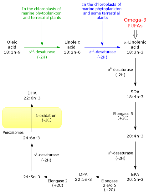
It should be emphasized that terrestrial vascular plants lack the enzymatic machinery necessary to synthesize omega-3 LC-PUFAs, such as EPA and DHA, the primary production of which is almost exclusively carried out by phytoplankton.[13]
Shared enzymes
The enzymes that catalyze the conversion of ALA to DHA are the same ones used in the metabolic pathways leading to the synthesis of omega-6, omega-7, and omega-9 PUFAs.[2]
Although Δ5-desaturase and Δ6-desaturase have omega-3 fatty acids as their preferred substrates, the Western diet exhibits a severely imbalanced LA/ALA ratio in favor of the former. Excess LA saturates the enzymes, thereby greatly reducing the conversion of ALA to EPA and DHA, although the synthesis of arachidonic acid (ARA) from LA also appears to be limited in humans.[16]
Finally, it should be noted that both the omega-3 and omega-6 families of polyunsaturated fatty acids exert a feedback inhibition on the biosynthesis of omega-9 polyunsaturated fatty acids.[4]
Biosynthesis of omega-3 fatty acids in humans
Many animals, including humans, possess the enzymatic capacity necessary to synthesize all other omega-3 fatty acids from dietary ALA.[3][4]
This metabolic pathway is primarily localized in the liver and within the cerebral microcirculation of the blood-brain barrier, including the cerebral endothelium and astrocytes, with limited overall efficiency and subject to high individual variability.[17][18]
Biosynthetic efficiency progressively decreases along the pathway: the conversion of ALA to EPA is limited, with Δ6-desaturase activity acting as the main limiting factor, while the synthesis of DHA from EPA is further reduced. It should also be noted that conversion efficiency is significantly higher in women of childbearing age than in men, due to the modulatory action of estrogens.[17]
However, recent genomic studies have demonstrated the existence of marked global polymorphism within the FADS gene cluster, located contiguously on chromosome 11q12.2, which encodes desaturases, specifically Δ5-desaturase, FADS1, and Δ6-desaturase, FADS2.[15]
A comparison of DNA extracted from Bronze Age human remains with that of contemporary populations has made it possible to trace the evolution of allele frequencies within the FADS cluster, highlighting how dietary adaptation has driven genotypic and phenotypic divergences.[19]
- In European populations, the transition from a hunter-gatherer society to an agricultural one led to an increase in LA and ALA intake and a decrease in EPA and ARA intake. Selective pressure would therefore have favored the haplotype associated with higher FADS1 expression and a concurrent downregulation of FADS2.[15]
- Among the Inuit of Greenland, an exactly opposite adaptive pattern has been observed. Intense natural selection has favored alleles that reduce the conversion of LA and ALA into LC-PUFAs, thereby compensating for the extremely high direct dietary intake of EPA and DHA.[20]
Conditionally essential fatty acids
The absence of Δ12-desaturase and Δ15-desaturase in mammals prevents the de novo synthesis of both LA and ALA, and consequently, of all omega-3 polyunsaturated fatty acids. Therefore, these two fatty acids are the only true essential fatty acids, while all omega-6 and omega-3 LC-PUFAs derived from them are defined as conditionally essential (or semi-essential), since their endogenous synthesis becomes insufficient to meet requirements in the absence of an adequate dietary intake of LA and ALA.[2][21]
Cellular and tissue distribution
Omega-3 polyunsaturated fatty acids, particularly EPA and DHA, are essential structural components of cell membranes, where they are esterified in phospholipids, primarily in phosphatidylethanolamine and, to a lesser extent, in phosphatidylcholine.[5]
Cell membranes are their primary storage site, particularly in muscle and nervous tissues.[11] For example, DHA is the major PUFA in the phospholipids of the cell membranes of the vertebrate nervous system and the photoreceptors of the retina.[22][23][24][25] Due to this structural characteristic, PUFAs, both omega-3 and omega-6, differ from most other fatty acids, which are primarily stored as triglycerides within adipose tissue.[1][4]
At the molecular level, in membrane phospholipids, PUFAs are esterified primarily at the sn-2 position, while the sn-1 position is occupied mainly by saturated fatty acids, particularly palmitic acid and stearic acid.[5]
EPA and DHA in other animals
Organisms lacking Δ15-desaturase are unable to synthesize ALA and, if necessary, must obtain it from the plants and/or animals they eat, and, where applicable, produce EPA and/or DHA.[2][10]
Terrestrial herbivorous vertebrates, such as cows, meet their needs for long-chain omega-3 polyunsaturated fatty acids by producing them from ALA obtained by eating the green parts of plants.[8]
There are animals that do not require EPA and DHA and are virtually devoid of them. Among these are terrestrial insects, in which only very low levels of EPA are found, comparable to those of vitamins. In these insects, EPA is synthesized from dietary ALA and is used for the production of eicosanoids involved in reproduction and the immune response.[26] In contrast, aquatic insects, such as species with aquatic metamorphosis belonging to the orders Odonata, Ephemeroptera, and Trichoptera, have high levels of EPA, while DHA is virtually absent.[27]
Several classes of phytoplankton, such as the Cryptophyceae and Dinophyceae, are very rich in EPA and DHA, while the Bacillariophyceae (diatoms) are very rich in EPA. In general, marine phytoplankton are the primary producers of EPA and DHA; therefore, aquatic ecosystems are their main source.[28][29] EPA and DHA move up the food chain, passing from invertebrates to fish and then to terrestrial animals, including humans.[26]
Omega-3 and human health
Omega-3 polyunsaturated fatty acids are essential components of a healthy, balanced diet, as they are necessary for development, beginning in fetal life, and are associated, throughout life, with improved health and a reduced risk of disease.[9]
Omega-3 PUFAs are essential for fetal neurological development, and their intake by pregnant women is particularly important during the third trimester of pregnancy, a phase marked by significant brain growth.[25]
Epidemiological studies and clinical trials have linked a high intake of EPA and DHA to a reduced risk of cardiovascular events and cardiac mortality. These effects are attributable to improvements in several risk factors: a marked dose-dependent reduction in blood triglycerides is observed, typically with doses of 2–4 g/day of EPA/DHA, a slight increase in HDL cholesterol, and a favorable modulation of inflammatory markers such as C-reactive protein, blood pressure, and heart rate, with a largely neutral effect on LDL cholesterol.[30][31]
Clinical trials on cardiovascular prevention using high-dose EPA formulations (4 g/day) have confirmed a significant reduction in coronary ischemic events in high-risk patients on statin therapy, although the administration of high doses of omega-3 requires careful clinical monitoring due to a dose-dependent increase in the risk of atrial fibrillation.[32][33]
EPA and DHA have also proven useful in the treatment of chronic inflammatory diseases, such as rheumatoid arthritis, with a potential adjunctive role in conditions such as asthma, psoriasis, and inflammatory bowel disease (IBD), driven primarily by the formation of specialized lipid mediators involved in inflammatory resolution.[34][35]
Omega-3 LC-PUFAs, on the other hand, appear to have minimal or no effect on the prevention and treatment of type 2 diabetes mellitus.[36]
Omega-3/omega-6 ratio
Numerous epidemiological studies suggest that a diet with a low omega-3/omega-6 ratio negatively affects health, contributing to the development of many chronic diseases.[9]
Lower rates of cancer, autoimmune diseases, and coronary heart disease have been observed in populations consuming a diet with a high omega-3/omega-6 ratio, particularly among the Inuit of Greenland, the Japanese, and other societies with high fish consumption.[37][38] However, the Western-style diet has gradually become richer in saturated fatty acids, poorer in omega-3 polyunsaturated fatty acids, and richer in omega-6 fatty acids, with an omega-3-to-omega-6 ratio ranging from 1:10 to 1:20, therefore far from the recommended ratio of 1:5.[34]
The low omega-3/omega-6 ratio is due to several factors, listed below.
- Plant-based foods are generally rich in ALA; however, in industrial agriculture, crops rich in omega-6 have been much more successful.[1]
- Low consumption of seafood and fish oil.[34]
- High consumption of animals raised on corn-based feed. In addition, the omega-3 content of some farmed fish species is lower than that of their wild counterparts.[2]
- High consumption of oils rich in omega-6 and low in omega-3, such as safflower, sunflower, soybean, and corn oils.[39]
It should be noted, however, that although the omega-3/omega-6 ratio retains epidemiological and educational value, current research and health organizations have shifted their focus to the total intake of EPA + DHA and the simultaneous reduction of excess LA, rather than solely on the numerical value of the ratio.[3][40][41]
Note: the omega-3/omega-6 ratio does not appear to influence either the prevention or treatment of type 2 diabetes.[36]
Molecular mechanisms of omega-3 PUFAs
The molecular mechanisms underlying the cellular and physiological effects of omega-3 polyunsaturated fatty acids, particularly EPA, DPA, and DHA, are gradually being elucidated. These mechanisms involve the incorporation of these fatty acids into membrane phospholipids, their release in reactions catalyzed by secretory phospholipase A2, and the subsequent enzymatic modifications that lead to the formation of bioactive lipid mediators, ranging from eicosanoids to SPMs (specialized pro-resolving mediators).[13]
Cell membranes
Omega-3 fatty acids are structural components of cell membranes, where they play an essential role in regulating membrane fluidity.[11] Thanks to this effect, omega-3 fatty acids modulate cell responses dependent on membrane proteins, a function that is particularly important in the eye, where the presence of DHA enables optimal rhodopsin activity.[24] The effect on membrane fluidity is also essential for poikilothermic animals, also known as “cold-blooded” animals, that live in cold waters or environments. In these animals, EPA and DHA, together with antifreeze proteins, help maintain the liquid-crystalline state of the membranes, preventing the freezing, or gelation, of membrane lipids, an effect known as homeoviscous adaptation.[42]
EPA and DHA are capable of modulating the formation of lipid rafts, microdomains with a specific lipid composition that can serve as platforms for receptor action and the initiation of intracellular signaling pathways.[43] By modulating their formation, they influence signaling pathways in different cell types, such as neurons, immune system cells, and cancer cells.[9] In this way, these two omega-3 fatty acids are able to modulate the activation of transcription factors, such as NF-κB, PPARs, and SREBPs, and, consequently, the patterns of gene expression regulated by them.[44] This role is central to their function in regulating fatty acid and triglyceride metabolism, inflammation, and adipocyte differentiation.[9]
SPM
EPA, DPA, and DHA, as well as ARA among the omega-6 fatty acids, are substrates for the production of numerous bioactive lipid mediators, including eicosanoids and SPMs, namely, maresins, D-series resolvins, and protectins, molecules with well-defined roles in the regulation of inflammation, immunity, platelet aggregation, renal function, and smooth muscle contraction.[45]
Eicosanoids produced from arachidonic acid, which, in terms of quantity, is the major substrate for their synthesis, play important physiological roles, but an excess of these compounds has been associated with numerous pathological processes.[9] An increase in the content of EPA and DHA in membrane phospholipids is accompanied by a reduction in ARA content, a reduction in the production of lipid mediators derived from ARA, and an increase in the production of lipid mediators derived from omega-3 fatty acids, specifically marine SPMs, protectins, and resolvins, those of the D series derived from DHA and those of the E series derived from EPA, molecules responsible for many of the anti-inflammatory, pro-resolving, and immunomodulatory actions attributed to omega-3 polyunsaturated fatty acids.[7]
Direct actions
EPA, DPA, and DHA can play a role in their free forms. In fact, they are capable of acting directly through G-protein-coupled membrane receptors, modulating their activity, or by binding to specific nuclear receptors, such as DHA binding to PPARα.[46]
Finally, they reduce the absorption of omega-6 polyunsaturated fatty acids, and at the enzymatic level, they competitively inhibit cyclooxygenase and lipoxygenase and compete for acyltransferases.[1]
Food sources
Fish from cold seas and aquatic invertebrates, namely crustaceans and mollusks, are the most important source of omega-3 polyunsaturated fatty acids for humans.[10] These animals are able to convert ALA into EPA, DPA, and DHA and, most importantly, to accumulate them along the marine food chain, which in this case begins with phytoplankton.[26] Furthermore, considering DHA alone, it is present in high concentrations in many fish oils, especially those from fish raised in cold waters. Note, however, that many of these oils are also rich in saturated fatty acids.[47]
For those who do not consume seafood, the livers of land animals, as well as game birds of the order Passeriformes, are good sources of EPA and DHA.[48][49]
As for plant sources, ALA is found in high concentrations in flaxseeds and flaxseed oil, walnuts, chia seeds, hemp seeds, and canola oil. Microalgae, such as Schizochytrium sp., represent the only direct plant-based source of EPA and DHA, used both in dietary supplements, as in the case of oil derived from Schizochytrium sp., which is extremely rich in DHA, and in the production of aquaculture feed.[50]
Regarding daily intakes, it is not yet clear what the recommended values are. The following table shows the values suggested by the FAO and EFSA, which have essentially been adopted in Italy by the SINU’s LARN guidelines.[3][41]
| Organization | Category | Recommendation |
|---|---|---|
| FAO | Adult men and non-pregnant or non-lactating adult women | At least 250 mg of EPA + DHA per day |
| Pregnant or lactating women | At least 300 mg of EPA + DHA per day, of which at least 200 mg should be DHA | |
| Children aged 2 to 4 years | 100–150 mg of EPA + DHA per day | |
| Children aged 4 to 6 years | 150–200 mg of EPA + DHA per day | |
| Children aged 6 to 10 years | 200–250 mg of EPA + DHA per day | |
| EFSA | Adult men and non-pregnant adult women | Adequate Intake (AI) is 250 mg of EPA + DHA per day |
| Pregnant or lactating women | An additional 100–200 mg of DHA per day | |
| Infants and young children aged 6 months to 2 years | 100 mg of DHA per day | |
| Children aged 2 to 18 years | “Considered consistent with adult recommendations” |
Omega-3 in foods
Polyunsaturated fatty acids in free form are particularly susceptible to oxidation caused by heating. The various processes that food undergoes during preparation could therefore reduce their content. However, since omega-3 polyunsaturated fatty acids are not present in foods in free form but are esterified within membrane phospholipids, they are much less susceptible to oxidative damage.[51]
When evaluating the EPA and DHA content in foods, expressing it as a percentage of total fatty acids rather than as an absolute quantitative value (mg/g of wet weight or edible portion) can lead to misinterpretation.
For example, consider a fatty fish such as salmon: its absolute EPA + DHA content is high, about 8 mg/g of wet weight, while expressing it as a percentage yields 20%.
In contrast, Atlantic cod has an EPA + DHA percentage of over 40%, but an absolute content of less than 3 mg/g of wet weight. In this case, the high percentage is an artifact resulting from the fact that cod is a lean fish with very few total lipids, whereas in fatty fish, the high absolute density of EPA + DHA appears to be “diluted” by the large reserve of other fatty acids, especially monounsaturated and saturated fatty acids, present in the animal’s adipose tissue.[52]
Conversely, if the EPA + DHA content is expressed in mg/g of product, no reduction is observed following most of the treatments to which the food is subjected during the preparation phase.[26]
References
- a b c d e f Akoh C.C., Min D.B. Food lipids: chemistry, nutrition, and biotechnology. 4th Edition. Boca Raton: CRC Press, 2017. doi:10.1201/9781315151854
- a b c d e f Chow C.K. Fatty acids in foods and their health implication. 3rd Edition. CRC Press, 2008. doi:10.1201/9781420006902
- a b c d EFSA Panel on Dietetic Products, Nutrition, and Allergies (NDA). Scientific opinion on dietary reference values for fats, including saturated fatty acids, polyunsaturated fatty acids, monounsaturated fatty acids, trans fatty acids, and cholesterol. 2010. doi:10.2903/j.efsa.2010.1461
- a b c d Sprecher H. Metabolism of highly unsaturated n-3 and n-6 fatty acids. Biochim Biophys Acta. 2000 Jul 19;1486(2-3):219-31. doi:10.1016/s1388-1981(00)00077-9
- a b c d Alberts B., Heald B., Johnson A.D., Morgan D., Raff M., Roberts K., Walter P. Molecular Biology of the Cell. 7th Edition. Garland Science, 2022.
- ^ Serhan C.N. Pro-resolving lipid mediators are leads for resolution physiology. Nature 2014;510(7503):92-101. doi:10.1038/nature13479
- a b Duvall M.G., Levy B.D. DHA- and EPA-derived resolvins, protectins, and maresins in airway inflammation. Eur J Pharmacol 2016;785:144-155. doi:10.1016/j.ejphar.2015.11.001
- a b Kouba M., Mourot J. A review of nutritional effects on fat composition of animal products with special emphasis on n-3 polyunsaturated fatty acids. Biochimie 2011;93(1):13-7. doi:10.1016/j.biochi.2010.02.027
- a b c d e f g Calder P.C. Very long-chain n-3 fatty acids and human health: fact, fiction and the future. Proc Nutr Soc 2018 77(1):52-72. doi:10.1017/S0029665117003950
- a b c d Tocher D.R., Betancor M.B., Sprague M., Olsen R.E., Napier J.A. Omega-3 long-chain polyunsaturated fatty acids, EPA and DHA: bridging the gap between supply and demand. Nutrients 2019;11(1):89. doi:10.3390/nu11010089
- a b c Stubbs C.D., Smith A.D. The modification of mammalian membrane polyunsaturated fatty acid composition in relation to membrane fluidity and function. Biochim Biophys Acta 1984;779(1):89-137. doi:10.1016/0304-4157(84)90005-4
- ^ Okuley J., Lightner J., Feldmann K., Yadav N., Lark E., Browse J. Arabidopsis FAD2 gene encodes the enzyme that is essential for polyunsaturated lipid synthesis. Plant Cell 1994;6(1):147-58. doi:10.1105/tpc.6.1.147
- a b c Lee J.M., Lee H., Kang S. and Park W.J. Fatty acid desaturases, polyunsaturated fatty acid regulation, and biotechnological advances. Nutrients 2016;8(1):23. doi:10.3390/nu8010023
- ^ Yadav N.S., Wierzbicki A., Aegerter M., Caster C.S., Pérez-Grau L., Kinney A.J., Hitz W.D., Booth J.R. Jr, Schweiger B., Stecca K.L., Allen S.M., Blackwell M., Reiter R.S., Carlson T.J., Russell S.H., Feldmann K.A., Pierce J., Browse J. Cloning of higher plant omega-3 fatty acid desaturases. Plant Physiol 1993;103(2):467-76. doi:10.1104/pp.103.2.467
- a b c Buckley M.T., Racimo F., Allentoft M.E., et al. Selection in Europeans on fatty acid desaturases associated with dietary changes. Mol Biol Evol 2017;34(6):1307-1318. doi:10.1093/molbev/msx103
- ^ Williams C.M., Burdge G. Long-chain n-3 PUFA: plant v. marine sources. Proc Nutr Soc 2006;65(1):42-50. doi:10.1079/pns2005473
- a b Burdge G.C., Wootton S.A. Conversion of α-linolenic acid to eicosapentaenoic, docosapentaenoic and docosahexaenoic acids in young women. Br J Nutr 2002;88(4):411-420. doi:10.1079/BJN2002689
- ^ Kim H-Y. Novel metabolism of docosahexaenoic acid in neural cells. J Biol Chem 2007;282(26):18661-18665. doi:10.1074/jbc.R700015200
- ^ Mathieson I., Lazaridis I., Rohland N., Mallick S., Patterson N., Roodenberg S.A., Harney E., Stewardson K., Fernandes D., Novak M., et al. Genome-wide patterns of selection in 230 ancient Eurasians. Nature 2015;528:499-503. doi:10.1038/nature16152
- ^ Fumagalli M., Moltke I., Grarup N., Racimo F., Bjerregaard P., Jørgensen M.E., Korneliussen T.S., Gerbault P., Skotte L., Linneberg A., Christensen C., Brandslund I., Jørgensen T., Huerta-Sánchez E., Schmidt E.B., Pedersen O., Hansen T., Albrechtsen A., Nielsen R.. Greenlandic Inuit show genetic signatures of diet and climate adaptation. Science 2015;349(6254):1343-7. doi:10.1126/science.aab2319
- ^ Smith W., Mukhopadhyay R. Essential fatty acids: the work of George and Mildred Burr. J Biol Chem 2012;287(42):35439-35441. doi:10.1074/jbc.O112.000005
- ^ Salem N. Jr, Litman B., Kim H.Y., Gawrisch K. Mechanisms of action of docosahexaenoic acid in the nervous system. Lipids 2001;36(9):945-59. doi:10.1007/s11745-001-0805-6
- ^ Lauritzen L., Hansen H.S., Jørgensen M.H., Michaelsen K.F. The essentiality of long chain n-3 fatty acids in relation to development and function of the brain and retina. Prog Lipid Res 2001;40(1-2):1-94. doi:10.1016/s0163-7827(00)00017-5
- a b Niu S.L., Mitchell D.C., Lim S.Y., Wen Z.M., Kim H.Y., Salem N. Jr, Litman B.J. Reduced G protein-coupled signaling efficiency in retinal rod outer segments in response to n-3 fatty acid deficiency. J Biol Chem 2004;279(30):31098-104. doi:10.1074/jbc.M404376200
- a b McCann J.C., Ames B.N. Is docosahexaenoic acid, an n-3 long-chain polyunsaturated fatty acid, required for development of normal brain function? An overview of evidence from cognitive and behavioral tests in humans and animals. Am J Clin Nutr 2005;82(2):281-95. doi:10.1093/ajcn.82.2.281
- a b c d Gladyshev M.I., Sushchik N.N. Long-chain omega-3 polyunsaturated fatty acids in natural ecosystems and the human diet: assumptions and challenges. Biomolecules 2019;9(9):485. doi:10.3390/biom9090485
- ^ Popova O.N., Haritonov A.Y., Sushchik N.N., Makhutova O.N., Kalachova G.S., Kolmakova A.A., Gladyshev M.I. Export of aquatic productivity, including highly unsaturated fatty acids, to terrestrial ecosystems via Odonata. Sci Total Environ 2017;581-582:40-48. doi:10.1016/j.scitotenv.2017.01.017
- ^ Ahlgren G., Lundstedt L., Brett M., Forsberg C. Lipid composition and food quality of some freshwater phytoplankton for cladoceran zooplankters. J Plankton Res 1990;12(4):809-18. doi:10.1093/plankt/12.4.809
- ^ Muller-Navarra D.C. Biochemical versus mineral limitation in Daphnia. Limnol Oceanogr 1995;40:1209-1214. doi:10.4319/lo.1995.40.7.1209
- ^ AbuMweis S., Jew S., Tayyem R. Agraib L. Eicosapentaenoic acid and docosahexaenoic acid containing supplements modulate risk factors for cardiovascular disease: a meta-analysis of randomised placebo-control human clinical. J Hum Nutr Diet 2018;31(1):67-84. doi:10.1111/jhn.12493
- ^ Mensink R.P., Zock P.L., Kester A.D., Katan M.B. Effects of dietary fatty acids and carbohydrates on the ratio of serum total to HDL cholesterol and on serum lipids and apolipoproteins: a meta-analysis of 60 controlled trials. Am J Clin Nutr 2003;77(5):1146-55. doi:10.1093/ajcn/77.5.1146
- ^ Bhatt D.L., Steg P.G., Miller M., Brinton E.A., Jacobson T.A., Ketchum S.B., Doyle R.T. Jr, Juliano R.A., Jiao L., Granowitz C., Tardif J.C., Ballantyne C.M; REDUCE-IT Investigators. Cardiovascular risk reduction with icosapent ethyl for hypertriglyceridemia. N Engl J Med 2019;380(1):11-22. doi:10.1056/NEJMoa1812792
- ^ Khan S.U., Lone A.N., Khan M.S., Virani S.S., Blumenthal R.S., Nasir K., Miller M., Michos E.D., Ballantyne C.M., Boden W.E., Bhatt D.L. Effect of omega-3 fatty acids on cardiovascular outcomes: a systematic review and meta-analysis. EClinicalMedicine 2021;38:100997. doi:10.1016/j.eclinm.2021.100997
- a b c Simopoulos A.P. The importance of the omega-6/omega-3 fatty acid ratio in cardiovascular disease and other chronic diseases. Exp Biol Med 2008;233(6):6746-88. doi:10.1016/S0753-3322(02)00253-6
- ^ Calder P.C. Omega-3 fatty acids and inflammatory processes: from molecules to man. Biochem Soc Trans 2017;45(5):1105-1115. doi:10.1042/BST20160474
- a b Brown T.J., Brainard J., Song F., Wang X., Abdelhamid A., Hooper L. Omega-3, omega-6, and total dietary polyunsaturated fat for prevention and treatment of type 2 diabetes mellitus: systematic review and meta-analysis of randomised controlled trials. BMJ 2019;366:l4697. doi:10.1136/bmj.l4697
- ^ Kromann N., Green A. Epidemiological studies in the Upernavik district, Greenland. Incidence of some chronic diseases 1950-1974. Acta Med Scand 1980;208(5):401-6. doi:10.1111/j.0954-6820.1980.tb01221.x
- ^ Yano K., MacLean C.J., Reed D.M., Shimizu Y., Sasaki H., Kodama K., Kato H., Kagan A. A comparison of the 12-year mortality and predictive factors of coronary heart disease among Japanese men in Japan and Hawaii. Am J Epidemiol 1988;127(3):476-87. doi:10.1093/oxfordjournals.aje.a114824
- ^ Blasbalg T.L., Hibbeln J.R., Ramsden C.E., Majchrzak S..F, Rawlings R.R. Changes in consumption of omega-3 and omega-6 fatty acids in the United States during the 20th century. Am J Clin Nutr 2011;93(5):950-62. doi:10.3945/ajcn.110.006643
- ^ SanGiovanni J.P., Chew E.Y. The role of omega-3 long-chain polyunsaturated fatty acids in health and disease of the retina. Prog Retin Eye Res 2005;24(1):87-138. doi:10.1016/j.preteyeres.2004.06.002
- a b FAO. Global recommendations for EPA and DHA intake (As of 30 June 2014). In: Fats and fatty acids in human nutrition. Report of an expert consultation. FAO Food and Nutrition Paper 91. Rome, 2010. URL: https://www.fao.org/fileadmin/user_upload/nutrition/docs/requirements/fatsandfattacidsreport.pdf
- ^ Hazel J.R. Thermal adaptation in biological membranes: is homeoviscous adaptation the explanation? Annu Rev Physiol 1995;57:19-42. doi:10.1146/annurev.ph.57.030195.000315
- ^ Wassall S.R., Stillwell W. Polyunsaturated fatty acid-cholesterol interactions: domain formation in membranes. Biochim Biophys Acta 2009;1788(1):24-32. doi:10.1016/j.bbamem.2008.10.011
- ^ Jump D.B., Botolin D., Wang Y., Xu J., Christian B., Demeure O. Fatty acid regulation of hepatic gene transcription. J Nutr 2005;135(11):2503-6. doi:10.1093/jn/135.11.2503
- ^ Kuda O. Bioactive metabolites of docosahexaenoic acid. Biochimie 2017;136:12-20. doi:10.1016/j.biochi.2017.01.002
- ^ Oh D.Y., Talukdar S., Bae E.J., Imamura T., Morinaga H., Fan WQ, Li P., Lu W.J., Watkins S.M., Olefsky J.M. GPR120 is an omega-3 fatty acid receptor mediating potent anti-inflammatory and insulin-sensitizing effects. Cell 2010 142(5):687-698. doi:10.1016/j.cell.2010.07.041
- ^ Mason R.P., Sherratt S.C.R. Omega-3 fatty acid fish oil dietary supplements contain saturated fats and oxidized lipids that may interfere with their intended biological benefits. Biochem Biophys Res Commun 2017;483(1):425-429. doi:10.1016/j.bbrc.2016.12.127
- ^ Gladyshev M.I., Makhutova O.N., Gubanenko G.A., Rechkina E.A., Kalachova G.S., Sushchik N.N. Livers of terrestrial production animals as a source of long-chain polyunsaturated fatty acids for humans: an alternative to fish? Eur J Lipid Sci Technol 2015;117:1417-1421. doi:10.1002/ejlt.201400449
- ^ Gladyshev M.I., Popova O.N., Makhutova, O.N., Zinchenko, T.D., Golovatyuk, L.V., Yurchenko, Y.A., Kalachova, G.S., Krylov, A.V., Sushchik, N.N. Comparison of fatty acid compositions in birds feeding in aquatic and terrestrial ecosystems. Contemp Probl Ecol 2016;9:503-513. doi:10.1134/S1995425516040065
- ^ U.S. Department of Agriculture, Agricultural Research Service. FoodData Central, 2019.
- ^ Belitz H.-D., Grosch W., Schieberle P. Food Chemistry. 4th Edition. Springer, 2009.
- ^ Gladyshev M.I., Sushchik N.N., Tolomeev A.P., Dgebuadze Y.Y. Meta-analysis of factors associated with omega-3 fatty acid contents of wild fish. Rev Fish Biol Fish 2018;28:277-299. doi:10.1007/s11160-017-9511-0
Domande Frequenti
Which omega-3 polyunsaturated fatty acids are essential for human health?
Key omega-3 polyunsaturated fatty acids include EPA, DHA, and ALA. EPA contains 20 carbon atoms and five double bonds, playing a critical role in cardiovascular protection, while DHA features 22 carbons and six double bonds, serving as a structural component in brain tissue.
How is ALA converted to long-chain omega-3 polyunsaturated fatty acids?
Among omega-3 polyunsaturated fatty acids, ALA is converted into EPA and DHA via hepatic desaturase and elongase enzymes. However, human metabolic conversion efficiency is physiologically limited, typically remaining below 5-10% for EPA synthesis and even less for DHA.
What is the recommended intake of omega-3 polyunsaturated fatty acids?
According to FAO and EFSA guidelines, the adequate intake of omega-3 polyunsaturated fatty acids (EPA + DHA) for healthy adults is at least 250 mg per day. Pregnancy and lactation require an additional 100-200 mg daily of DHA to support optimal fetal development.