

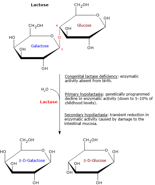
Hypolactasia, or lactase deficiency, is a biochemical condition characterized by reduced activity of lactase, the enzyme responsible for hydrolyzing lactose into its constituent monosaccharides, glucose and galactose, which are then absorbed into the bloodstream.[1][2]
When lactase activity is deficient or absent, unabsorbed lactose remains in the intestinal lumen, leading to lactose malabsorption. As undigested lactose travels downstream into the colon, it becomes an osmotic substrate and an energetic source for the gut microbiota, triggering a cascade of processes that can give rise to gastrointestinal symptoms, a clinical condition defined as lactose intolerance.[3][4]
Etiologically, hypolactasia can be classified based on its underlying causes into primary hypolactasia and congenital lactase deficiency, which have genetic basis, and secondary hypolactasia, a transient and not genetically programmed condition.[1]
Summary: Key Points
- Definition and mechanisms: hypolactasia is a biochemical condition characterized by reduced lactase activity in the small intestine, leading to lactose malabsorption and potential colonic fermentation by gut microbiota.
- Clinical manifestations: symptoms of lactose intolerance include abdominal pain, bloating, flatulence, and osmotic diarrhea, which occur in only 30–50% of hypolactasic individuals depending on physiological modulating factors.
- Congenital lactase deficiency: an extremely rare, life-threatening genetic disorder present at birth.
- Primary hypolactasia: a genetically programmed, post-weaning decline in lactase activity that represents the phylogenetically normal phenotype in roughly three-quarters of the human population, with geographic and ethnic variability.
- Secondary hypolactasia: a transient reduction in lactase activity caused by mucosal damage.
Contents
- Undigested lactose and gut microbiota
- From hypolactasia to lactose intolerance
- Congenital lactose intolerance
- Primary hypolactasia
- Secondary hypolactasia
- References
Undigested lactose and gut microbiota
In the colon, the disaccharide can be partially fermented by gut microbiota bacteria, producing an excess of gas such as molecular nitrogen and hydrogen, carbon dioxide, methane, and short-chain fatty acids, such as butyric acid. In addition, fermentation products and undigested carbohydrates act as osmotically active solutes, increasing intraluminal osmotic pressure and water content in the colon.[5][6]
The symptoms that characterize lactose intolerance, resulting from the processes described above, include:
- abdominal pain, due to the acidification of the colon contents and to an irritating effect on the mucosa resulting from excess short-chain fatty acids;
- flatulence, bloating and abdominal distension, caused by excessive gas production;
- diarrhea, as a result of the increased volume of water in the colon due to the presence of osmotically active solutes and to the reduction in the efficiency of water absorption due to the increased motor activity of the colon induced by excess short-chain fatty acids.[3]
It should be noted that the cascade of events described above is applicable to any sugar that is not absorbed in the small intestine, as well as excessive dietary fiber intake.
From hypolactasia to lactose intolerance
Hypolactasia has only been recognized in the last 50 years and is the most common food malabsorption condition. However, it is not always accompanied by lactose intolerance, with symptoms manifesting in only 30–50% of cases.[1] While residual lactase activity is a key factor, several other physiological variables modulate symptom onset.
- Amount of lactose ingested, as the risk increases as the amount of the disaccharide increases.
- Gastric emptying time, which is influenced by the composition of the meal. For example, lipids, proteins, and fibers can effectively slow down the emptying, allowing the disaccharide to enter the small intestine slowly.
- Intestinal transit time, which influences the contact time between the enzyme and lactose. Therefore, foods that slow down transit time may increase the probability of contact.
- Colonic microbiota composition, which may affect fermentation processes.[7]

Congenital lactose intolerance
Congenital lactose intolerance, a disease inherited in an autosomal recessive pattern, is an extremely rare life-threatening inborn error of metabolism.[8] Lactase activity is undetectable, and affected newborns present with severe diarrhea within the first days of life. The only effective treatment is a strict lactose-free diet, which eliminates symptoms and enables normal growth and development.[4]
Primary hypolactasia
In approximately three-quarters of the world’s population, as in most mammals, a genetically programmed age-dependent decline in lactase activity occurs after weaning. This reduction is not due to damage to the gene encoding this protein, located on chromosome 2, but is genetically programmed and inherited in an autosomal recessive pattern. Residual lactase activity can be as low as 5–10% of that present during infancy.[1][8]
Geographical and ethnic distribution
The prevalence of primary adult hypolactasia exhibits striking geographic and ethnic variability, largely shaped by the ancestral presence of pastoralist culture and dairy farming.
In populations with a long tradition of pastoralism, such as Northern Europeans and certain nomadic groups in Arabia and Africa, lactase persistence is the norm, and primary hypolactasia is extremely rare. Conversely, in most Mediterranean, African, and Asian populations, lactase activity declines rapidly after weaning, reaching near-universal levels of hypolactasia in countries such as Thailand or among the Yoruba people of West Africa.[9][10]
| Population/Geographic region | Hypolactasia prevalence (%) | Lactase persistence (%) |
|---|---|---|
| Global human population (estimated mean) | ≈ 65–75% | ≈ 25–35% |
| Northern Europe (e.g., Denmark, Sweden) | 3–5% | 95–97% |
| White north americans | 5–20% | 80–95% |
| Mediterranean region and southern Europe | 50–70% | 30–50% |
| Black north americans | 70–75% | 25–30% |
| Asian and filipino populations | 85–95% | 5–15% |
| Thai population | up to 99% | ≈ 1% |
| Yoruba Ethnic groups (Nigeria, Benin, Togo) | up to 99% | ≈ 1% |
| Nomadic pastoralist populations (arid zones of Arabia and North Africa) | Low (< 20%) | High (> 80%) |
Therefore, primary hypolactasia is not a disease, but should be considered phylogenetically normal and is not affected by continuous exposure to milk or lactose.[4]
In primary hypolactasia, the reduction of lactase activity is not uniformly distributed throughout the tissue. Instead, it exhibits a patchy pattern, even within the same villus, between a predominant number of non-lactase-producing enterocytes and a smaller population of enterocytes that produce high amounts of the enzyme.[11]
Evolutionary advantage
The persistence of lactase activity likely provided an evolutionary survival advantage. With the emergence of dairy farming approximately 10,000 years ago, milk consumption provided individuals with sufficient lactase activity a distinct survival advantage during severe winters in Northern Europe and during periods of crop failure.[7]
Secondary hypolactasia
Secondary hypolactasia results from conditions causing structural damage to enterocytes such as intestinal infections, cow’s milk protein allergy, Crohn’s disease, celiac disease, an autoimmune reaction to dietary gluten, drug therapies, surgery, radiation to the gastrointestinal tract, or excessive alcohol consumption. Under these circumstances, many brush border proteins are affected, and among the glycosidases, lactase undergoes the most marked reduction.[12]
Secondary hypolactasia is most common in developing nations where chronic gastrointestinal infections are prevalent.
Once the underlying cause is resolved, the intestinal epithelium heals, lactase activity increases, and it eventually returns to baseline levels. Complete recovery can take up to six months, during which avoiding milk and lactose-containing foods is recommended.[4]
References
- a b c d Fassio F., Facioni M.S., Guagnini F. Lactose maldigestion, malabsorption, and intolerance: a comprehensive review with a focus on current management and future perspectives. Nutrients 2018;10(11):1599. doi:10.3390/nu10111599
- ^ Troelsen J.T. Adult-type hypolactasia and regulation of lactase expression. Biochim Biophys Acta 2005;1723(1-3):19-32. doi:10.1016/j.bbagen.2005.02.003
- a b Misselwitz B., Butter M., Verbeke K., Fox M.R. Update on lactose malabsorption and intolerance: pathogenesis, diagnosis and clinical management. Gut 2019;68(11):2080-2091. doi:10.1136/gutjnl-2019-318404
- a b c d Suarez F., Shannon C., Hertzler S., Savaiano D. Food intolerance | Lactose Intolerance. Editor(s): Caballero B. Encyclopedia of food sciences and nutrition. 2nd Edition. Academic Press, 2003;2634-2642. doi:10.1016/B0-12-227055-X/00511-3
- ^ Holtug K., Clausen M.R., Hove H., Christiansen J., Mortensen P.B. The colon in carbohydrate malabsorption: short-chain fatty acids, pH, and osmotic diarrhoea. Scand J Gastroenterol 1992;27(7):545-52. doi:10.3109/00365529209000118
- ^ Weijers H.A., va de Kamer J.H., Mossel D.A., Dicke W.K. Diarrhoea caused by deficiency of sugar-splitting enzymes. Lancet 1960;2(7145):296-7. doi:10.1016/s0140-6736(60)91381-7
- a b Di Stefano M. Il malassorbimento e l’intolleranza al lattosio. Fisiopatologia, diagnosi e approccio terapeutico. SIMG 2012;5:40-45.
- a b Heyman M.B.; Committee on Nutrition. Lactose intolerance in infants, children, and adolescents. Pediatrics 2006;118(3):1279-86. doi:10.1542/peds.2006-1721
- ^ Swallow D.M. Genetics of lactase persistence and lactose intolerance. Annu Rev Genet 2003;37:197-219. doi:10.1146/annurev.genet.37.110801.143820
- ^ Misselwitz B., Pohl D., Frühauf H., Fried M., Vavricka S.R., Fox M. Lactose malabsorption and intolerance: pathogenesis, diagnosis and treatment. United European Gastroenterol J 2013;1(3):151-9. doi:10.1177/2050640613484463
- ^ Majuri L., Raja V., Potter J., Swallow D., Wan Ho M., Fiocca R., Finzi G., Cornaggia M., Capella C., Quaroni A., Auricchio A. Mosaic pattern of lactase expression by villus enterocytes in human adult-type hypolactasia. Pediatr Res 1990;27:532. doi:10.1203/00006450-199005000-00040
- ^ Lomer M.C., Parkes G.C., Sanderson J.D. Review article: lactose intolerance in clinical practice – myths and realities. Aliment Pharmacol Ther 2008;27(2):93-103. doi:10.1111/j.1365-2036.2007.03557.x