

Multienzyme complexes are discrete and stable structures composed of noncovalently associated enzymes that catalyze two or more sequential steps of a metabolic pathway.[1]
They can be considered a step forward in the evolution of catalytic efficiency, as they provide advantages that individual enzymes, even those that have achieved catalytic perfection, would not have on their own.[2]
Contents
- The advantages of multienzyme complexes
- How metabolic channeling optimizes biochemical reactions
- Examples of multienzyme complexes
- References
The advantages of multienzyme complexes
During evolution, some enzymes have reached a state of virtual catalytic perfection, meaning that nearly every collision with their substrate results in catalysis. Below are several noteworthy examples.
- Fumarase (EC 4.2.1.2) catalyzes the seventh reaction of the citric acid cycle, the reversible hydration/dehydration of fumarate’s double bond to form malate.
- Acetylcholinesterase (EC 3.1.1.7) catalyzes the hydrolysis of acetylcholine, a neurotransmitter, into choline and acetic acid, which then dissociates to form a hydrogen ion and acetate.
- Superoxide dismutase (EC 1.15.1.1) catalyzes the conversion and inactivation of the highly reactive superoxide radical (O2.-) into hydrogen peroxide (H2O2)and water.
- Catalase (EC 1.11.1.6) catalyzes the degradation of H2O2 into water and oxygen.[2]
The rate of an enzymatic reaction is partly determined by how often enzymes and substrates collide. A simple way to increase this rate would be to increase enzyme and substrate concentrations. However, within a cell, their concentrations cannot be very high due to the large number of different reactions occurring simultaneously. In fact, most reactants are present in micromolar concentrations (10-6 M), while most enzymes are found at even lower concentrations.[3]
How metabolic channeling optimizes biochemical reactions
To overcome these limitations, evolution has taken different routes to enhance reaction rates. One such route is the optimization of enzyme spatial organization through the formation of multienzyme complexes and multifunctional enzymes. These structures minimize the distance that a reaction product must travel to reach the next active site, which is located close by.[1]
In other words, this leads to substrate (or metabolic) channeling, which can also occur through intramolecular channels connecting active sites.[4]
Metabolic channeling increases not just reaction rate but overall catalytic efficiency in several ways:
- minimizing diffusion of substrates and products into bulk solvent, which prevents dilution and maintains high local concentrations, even when overall intracellular concentrations are low;
- reducing the time required for substrates to diffuse between successive active sites;
- lowering the probability of side reactions;
- protecting chemically labile intermediates from degradation by the solvent.[2][4]
Another metabolic advantage, similar to multifunctional enzymes, is that multienzyme complexes allow coordinated regulation of the catalytic activity of their constituent enzymes. Since the enzyme catalyzing the first reaction in a pathway is often the regulatory enzyme, this prevents:
- the synthesis of unneeded intermediates that would occur if regulation happened downstream;
- the removal of metabolites from other pathways, as well as energy waste.[2]
Examples of multienzyme complexes
As noted above, especially in eukaryotes, multienzyme complexes, like multifunctional enzymes, are common in both anabolic and catabolic pathways, whereas few enzymes diffuse freely in solution. Two examples of multienzyme complexes include the tryptophan synthase complex (EC 4.2.1.20), whose tunnel was the first to be discovered, and bacterial carbamoyl phosphate synthase complex (EC 6.3.4.16).[5][6][7]
2-Ketoacid dehydrogenase family
A classic case of multienzyme organization is the 2-ketoacid (or 2-oxoacid) dehydrogenase family, which includes:
- pyruvate dehydrogenase complex (PDC);
- branched-chain α-keto acid dehydrogenase complex (BCKDH);
- α-ketoglutarate dehydrogenase complex, also called 2-oxoglutarate dehydrogenase (OGDH).[1][8]

These multienzyme complexes are structurally and functionally similar. PDC, for instance, consists of multiple copies of three enzymes:
- pyruvate dehydrogenase (E1; EC 1.2.4.1);
- dihydrolipoyl transacetylase (E2; EC 2.3.1.12);
- dihydrolipoyl dehydrogenase (E3; EC 1.8.1.4).
The same E1–E2–E3 architecture occurs in all three complexes, both in prokaryotes and eukaryotes. Within a given species, E3 is identical, while E1 and E2 are homologous.[9]
Though substrate-specific, these enzymes share the same cofactors: coenzyme A, NAD+, thiamine pyrophosphate, FAD, and lipoamide. For clarity, their components are labeled:
- E1p, E1o and E1b (EC 1.2.4.4);
- E3p, E3o, and E3b (EC 1.8.1.4).[2]
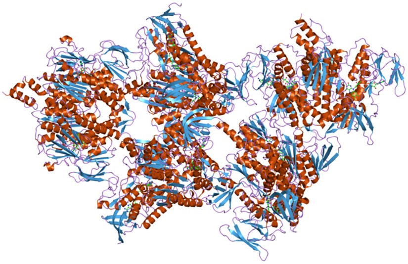
Note: eukaryotic PDC is the largest known multienzyme complex, larger than a ribosome, and can be visualized by electron microscopy.[10]
PDC links glycolysis to the citric acid cycle, catalyzing the irreversible oxidative decarboxylation of pyruvate to acetyl-CoA, with carbon dioxide (CO2) release and electron transfer to NAD+. Similarly, OGDH and BCKDH catalyze oxidative decarboxylations of α-ketoglutarate (to succinyl-CoA) and branched-chain α-keto acids (from valine, leucine, and isoleucine), involving:
- CO2 release;
- acyl group transfer to coenzyme A (forming acyl-CoA derivatives);
- NAD+ reduction to NADH.
The striking similarities in structure, cofactors, and mechanisms point to a common evolutionary origin.[10]
Tryptophan synthase complex
The tryptophan synthase complex is one of the best-studied examples of substrate channeling.[4] It is present in bacteria and plants, but not in animals.[11] In bacteria, it is composed of two α and two β subunits that associate into αβ dimers, which represent the functional units of the complex. These dimers further assemble into an αββα tetramer.[5][6]
The complex catalyzes the final two steps of tryptophan biosynthesis. In the first step, indole-3-glycerol phosphate undergoes an aldol cleavage, catalyzed by a lyase (EC 4.1.2.8) located on the α subunits, producing indole and glyceraldehyde 3-phosphate. Indole then reaches the active site of the β subunit through an approximately 30 Å-long hydrophobic tunnel that, within each αβ dimer, connects the two active sites. In the second step, in the presence of pyridoxal 5-phosphate, indole condenses with serine to form tryptophan.[4][12]
Acetyl-CoA carboxylase
Acetyl-CoA carboxylase (ACC; EC 6.4.1.2), a member of the biotin-dependent carboxylase family, catalyzes the committed step of fatty acid synthesis: the carboxylation of acetyl-CoA to malonyl-CoA. Malonyl-CoA, in turn, serves as a donor of two-carbon units for the elongation process leading to the synthesis of palmitic acid, catalyzed by fatty acid synthase (EC 2.3.1.85).[13]
In bacteria, ACC is a multienzyme complex composed of two enzymes, biotin carboxylase (EC 6.3.4.14) and carboxytransferase, plus a biotin carboxyl-carrier protein (BCCP).[13]
Conversely, in mammals and birds, it is a multifunctional enzyme, with both enzymatic activities and BCCP present on the same polypeptide chain.[8]
In higher plants, both forms are present.[14]
Carbamoyl phosphate synthetase complex
Another well-characterized example of substrate channeling is the bacterial carbamoyl phosphate synthetase complex, which catalyzes the synthesis of carbamoyl phosphate, required for pyrimidine and arginine synthesis. This multienzyme complex contains an approximately 100 Å-long tunnel that connects its three active sites.
The first active site catalyzes the release of the amide nitrogen from glutamine as an ammonium ion, which enters the tunnel and reaches the second active site, where it is combined with bicarbonate, at the expense of ATP, to yield carbamate. In the last active site, carbamate is phosphorylated to form carbamoyl phosphate.[7][15]
References
- ^ a b c Michal G., Schomburg D. Biochemical pathways. An atlas of biochemistry and molecular biology. 2nd Edition. John Wiley J. & Sons, Inc. 2012.
- ^ a b c d e Voet D. and Voet J.D. Biochemistry. 4th Edition. John Wiley J. & Sons, Inc. 2011.
- ^ Alberts B., Johnson A., Lewis J., Morgan D., Raff M., Roberts K., Walter P. Molecular biology of the cell. 6th Edition. Garland Science, Taylor & Francis Group, 2015.
- ^ a b c d Moran L.A., Horton H.R., Scrimgeour K.G., Perry M.D. Principles of Biochemistry. 5th Edition. Pearson, 2012.
- ^ a b Hyde C.C., Ahmed S.A., Padlan E.A., Miles E.W., and Davies D.R. Three-dimensional structure of the tryptophan synthase multienzyme complex from Salmonella typhimurium. J Biol Chem 1988;263(33):17857-17871. doi:10.1016/S0021-9258(19)77913-7
- ^ a b Hyde C.C., Miles E.W. The tryptophan synthase multienzyme complex: exploring structure-function relationships with X-ray crystallography and mutagenesis. Nat Biotechnol 1990:8;27-32. doi:10.1038/nbt0190-27
- ^ a b Thoden J.B., Holden H.M., Wesenberg G., Raushel F.M., Rayment I. Structure of carbamoyl phosphate synthetase: a journey of 96 A from substrate to product. Biochemistry 1997;36(21):6305-16. doi:10.1021/bi970503q
- ^ a b Garrett R.H., Grisham C.M. Biochemistry. 4th Edition. Brooks/Cole, Cengage Learning, 2010.
- ^ Zhou Z.H., McCarthy D.B., O’Connor C.M., Reed L.J., and J.K. Stoops. The remarkable structural and functional organization of the eukaryotic pyruvate dehydrogenase complexes. Proc Natl Acad Sci USA 2001;98(26):14802-14807. doi:10.1073/pnas.011597698
- ^ a b Nelson D.L., Cox M.M. Lehninger. Principles of biochemistry. 8th Edition. W.H. Freeman and Company, 2021.
- ^ Xie G., Forst C., Bonner C., Jensen R.A. Significance of two distinct types of tryptophan synthase beta chain in Bacteria, Archaea and higher plants. Genome Biol 2002;3(1):RESEARCH0004. doi:10.1186/gb-2001-3-1-research0004
- ^ Hilario E., Caulkins B.G., Huang Y-M. M., You W., Chang C-E.A., Mueller L.J., Dunn M.F., and Fan L. Visualizing the tunnel in tryptophan synthase with crystallography: insights into a selective filter for accommodating indole and rejecting water. Biochim Biophys Acta 2016;1864(3):268-279. doi:10.1016/j.bbapap.2015.12.006
- ^ a b Cronan J.E. Jr, Waldrop G.L. Multi-subunit acetyl-CoA carboxylases. Prog Lipid Res 2002;41(5):407-35. doi:10.1016/s0163-7827(02)00007-3
- ^ Sasaki Y., Nagano Y. Plant acetyl-CoA carboxylase: structure, biosynthesis, regulation, and gene manipulation for plant breeding. Biosci Biotechnol Biochem 2004;68(6):1175-84. doi:10.1271/bbb.68.1175
- ^ Holden H.M., Thoden J.B., Raushel F.M. Carbamoyl phosphate synthetase: an amazing biochemical odyssey from substrate to product. Cell Mol Life Sci 1999;56(5-6):507-22. doi:10.1007/s000180050448